

Hip
Hip Joint
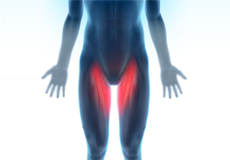
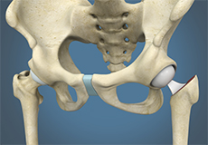
The hip joint is the largest weight-bearing joint in the human body. It is also referred to as a ball and socket joint and is surrounded by muscles, ligaments, and tendons. The thigh bone or femur and the pelvis join to form the hip joint.
Any injury or disease of the hip will adversely affect the joint's range of motion and ability to bear weight.
The hip joint is made up of the following:
- Bones and joints
- Ligaments of the joint capsule
- Muscles and tendons
- Nerves and blood vessels that supply the bones and muscles of the hip
Bones and Joints
The hip joint is the junction where the hip joins the leg to the trunk of the body. It is comprised of two bones: the thigh bone or femur and the pelvis which is made up of three bones called ilium, ischium, and pubis. The ball of the hip joint is made by the femoral head while the socket is formed by the acetabulum. The Acetabulum is a deep, circular socket formed on the outer edge of the pelvis by the union of three bones: ilium, ischium, and pubis. The lower part of the ilium is attached by the pubis while the ischium is considerably behind the pubis. The stability of the hip is provided by the joint capsule or acetabulum and the muscles and ligaments which surround and support the hip joint.
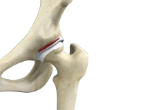
The head of the femur rotates and glides within the acetabulum. A fibrocartilagenous lining called the labrum is attached to the acetabulum and further increases the depth of the socket.
The femur or thigh bone is one of the longest bones in the human body. The upper part of the thigh bone consists of the femoral head, femoral neck, and greater and lesser trochanters. The head of the femur joins the pelvis (acetabulum) to form the hip joint. Next, to the femoral neck, there are two protrusions known as greater and lesser trochanters which serve as sites of muscle attachment.
Articular cartilage is the thin, tough, flexible, and slippery surface lubricated by synovial fluid that covers the weight-bearing bones of the body. It enables smooth movements of the bones and reduces friction.
Ligaments
Ligaments are fibrous structures that connect bones to other bones. The hip joint is encircled with ligaments to provide stability to the hip by forming a dense and fibrous structure around the joint capsule. The ligaments adjoining the hip joint include:
- Iliofemoral ligament: This is a Y-shaped ligament that connects the pelvis to the femoral head at the front of the joint. It helps in limiting the over-extension of the hip.
- Pubofemoral ligament: This is a triangular shaped ligament that extends between the upper portion of the pubis and the iliofemoral ligament. It attaches the pubis to the femoral head.
- Ischiofemoral ligament: This is a group of strong fibers that arise from the ischium behind the acetabulum and merge with the fibers of the joint capsule.
- Ligamentum teres: This is a small ligament that extends from the tip of the femoral head to the acetabulum. Although it has no role in hip movement, it does have a small artery within that supplies blood to a part of the femoral head.
- Acetabular labrum: The labrum is a fibrous cartilage ring which lines the acetabular socket. It deepens the cavity, increasing the stability and strength of the hip joint.
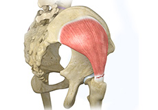
Muscles and Tendons
A long tendon called the iliotibial band runs along the femur from the hip to the knee and serves as an attachment site for several hip muscles including the following:
- Gluteals: These are the muscles that form the buttocks. There are three muscles (gluteus minimus, gluteus maximus, and gluteus medius) that attach to the back of the pelvis and insert into the greater trochanter of the femur.
- Adductors: These muscles are located in the thigh which helps in adduction, the action of pulling the leg back towards the midline.
- Iliopsoas: This muscle is located in front of the hip joint and provides flexion. It is a deep muscle that originates from the lower back and pelvis and extends up to the inside surface of the upper part of the femur.
- Rectus femoris: This is the largest band of muscles located in front of the thigh. They also are hip flexors.
- Hamstring muscles: These begin at the bottom of the pelvis and run down the back of the thigh. Because they cross the back of the hip joint, they help in extension of the hip by pulling it backward.
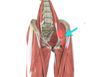
Nerves and Arteries
Nerves of the hip transfer signals from the brain to the muscles to aid in hip movement. They also carry the sensory signals such as touch, pain, and temperature back to the brain.
- The main nerves in the hip region include the femoral nerve in the front of the femur and the sciatic nerve at the back. The hip is also supplied by a smaller nerve known as the obturator nerve.
- In addition to these nerves, there are blood vessels that supply blood to the lower limbs. The femoral artery, one of the largest arteries in the body, arises deep in the pelvis and can be felt in front of the upper thigh.
Hip Movements
All of the anatomical parts of the hip work together to enable various hip movements. Hip movements include flexion, extension, abduction, adduction, circumduction, and hip rotation.
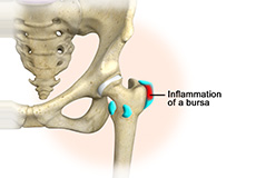
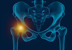
Hip Bursitis
Hip bursitis is a painful condition caused by inflammation of a bursa in the hip. Bursae are fluid- filled sacs present in joints between bone and soft tissue to reduce friction and provide cushioning during movement.
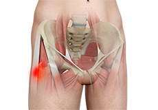
Hip Injury
The hip joint is one of the most important and flexible joints in the human body which allows us to walk, run, bend and perform physical activities. It is a ball (femoral head) and socket joint formed between the hip bone and femur (thighbone). It is surrounded by strong muscles and tough ligaments that prevent its dislocation.
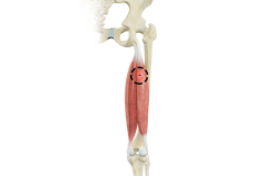
Hamstring Injuries
The hamstring is a group of three muscles that run along the back of the thigh from the hip to the knee. Hamstring injuries occur when these muscles are strained or pulled. They are common in dancers and athletes of all sorts including runners and those who play football, soccer, basketball, tennis, etc.
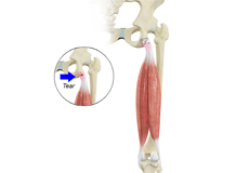
Gluteus Tendon Tear
The gluteal muscles (situated in the buttocks) are necessary for the stability and movement of the hip joints. The tendons of two gluteal muscles (gluteus medius and gluteal minimus) are attached at the outer hip region and are often called the “rotator cuff of the hip.”
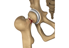
Femoroacetabular Impingement
Femoroacetabular impingement (FAI) is a condition characterized by excessive friction in the hip joint from the presence of bony irregularities. These cause pain and decreased range of hip motion.
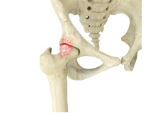
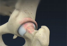
Hip Labral Tear
A hip labral tear is an injury to the labrum, the cartilage that surrounds the outside rim of your hip joint socket.
Hip Abductor Tears
Hip abductors are a major group of muscles found in the buttocks. It includes the gluteus maximus, gluteus medius, gluteus minimus, and tensor fascia lata muscles.
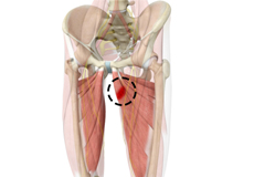
Hip Adductor Injuries
A hip adductor injury is also called a groin strain or groin tear and involves any of the adductor muscles. These injuries are common among athletes and those who play hockey, football, soccer, basketball, tennis, baseball, etc.
Hip Flexor Strain
A hip flexor strain is an overuse injury to the flexor muscles of your hip and can range from a minor stretch injury to a complete tear of the muscle fibers or tendons.
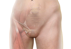
Hip Groin Disorders
The rehabilitation time for hip and groin injuries is longer than most other injuries, therefore, early and accurate diagnosis is essential. The management of hip and groin injuries is complex due to the presence of multiple anatomic structures in that region.
Hip Tendonitis
Tendons are strong connective tissue structures that connect muscle to bone. Hip tendonitis is a condition associated with degeneration of the hip tendons. This condition is mainly caused due to strain on the tendons which may occur due to overuse or biomechanical problems.
Groin Injuries in Athletes
Groin injuries are injuries sustained by athletes during sports activity. Groin injuries comprise about 2 to 5 percent of all sports injuries. The most common kind of groin injury is a groin strain or a pulled groin muscle.
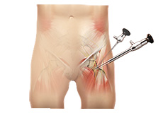
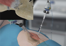
Hip Arthroscopy
Arthroscopy, also referred to as keyhole or minimally invasive surgery, is a procedure in which an arthroscope is inserted into a joint to check for any damage and repair it simultaneously.
Hip Arthroscopy - Supine Position
Arthroscopy, also referred to as keyhole or minimally invasive surgery, is a procedure in which an arthroscope is inserted into a joint to check for any damage and to repair the problem at the same time.
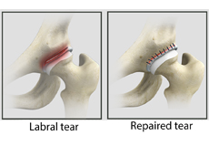
Hip Labral Repair
Labrum is a ring of strong fibrocartilaginous tissue lining around the socket of the hip joint. Labrum serves many functions where it acts as a shock absorber, lubricates the joint, and distributes the pressure equally.
Proximal Hamstring Repair
Hamstring injuries primarily occur when the muscle is exposed to extreme strain; when it is stretched beyond its ability or when it must withstand a sudden load.
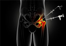
Femoroacetabular Osteoplasty
Femoroacetabular osteoplasty is the surgical reshaping of the protruding bony surface of the femur or acetabulum of the hip joint. FAO is performed arthroscopically as a minimally invasive procedure. An arthroscope is a small, fiber-optic instrument consisting of a lens, light source, and video camera.
Hip Fracture ORIF
A hip fracture is a break that occurs near the hip in the upper part of the femur or thighbone. The thighbone has two bony processes on the upper part - the greater and lesser trochanters. The lesser trochanter projects from the base of the femoral neck on the back of the thighbone.
Arthroscopic Gluteus Medius Tendon Repair
Arthroscopic gluteus medius tendon repair is a minimally invasive surgical procedure employed for the treatment of a gluteus medius tendon tear, when the tear does not respond to conservative treatment.
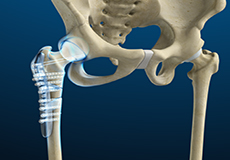
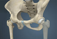
Hip Hemiarthroplasty
Hip hemiarthroplasty is a surgical technique employed to treat hip fractures. In this procedure, only one half (ball section) of the hip joint is substituted by a metal prosthesis.
Hip Fracture Surgery
Hip fractures involve a break that occurs near the hip in the upper part of the femur or thigh bone. The thigh bone has two bony processes on the upper part - the greater and lesser trochanters. The lesser trochanter projects from the base of the femoral neck on the back of the thigh bone.
Physical Therapy for Hip
Physical therapy is an exercise program that helps you to improve movement, relieve pain, encourage blood flow for faster healing, and restore your physical function and fitness level. The main aim of physical therapy is to make your daily activities, such as walking, getting in and out of bed and climbing stairs, easier. It can be prescribed as an individual treatment program or combined with other treatments.
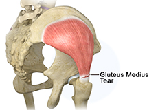
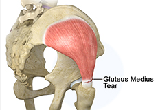
Gluteus Medius Tear
The gluteus medius is one of the major muscles of the hip; essential for the movement of the lower body and keeping the pelvis level during ambulation. The gluteus medius muscle arises from the top of the pelvic bone and attaches to the outer side of the thighbone or femur at the greater trochanter by the gluteus medius tendon.

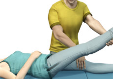
Physical Examination of the Hip
The hip joint is a ball and socket joint. The ball is the head of the femur (thighbone) that fits in the acetabulum (the hip socket). Tendons, muscles, and ligaments hold the joint in place. Articular cartilage covers the acetabulum and the femoral head. The synovial membrane of the hip joint secretes the synovial fluid which lubricates the joint enabling smooth movement.